X-ray is an electromagnetic
radiation of extremely short wavelength and high frequency between the gamma
and the ultraviolet radiation. These are commonly produced by accelerating (or
decelerating) charged particles using high-voltage electricity supply at a
piece of metal (typically tungsten). What gets reflected beck is X-rays. And X-rays
are the oldest sources of EM radiation used for imaging.
figure 1:
Producing X-rays
X-rays are roughly classified into two types
1. Soft x-rays
These have different optical
properties than visible light and therefore experiments must take place in
ultra high vacuum, where the photon beam is manipulated using special mirrors
and diffraction gratings.
2. Hard x-rays
These are the highest energy
x-rays, while the lower energy x-rays are referred to as soft x-rays. The
distinction between hard and soft x-rays is not well defined. Hard x-rays are
typically those with energies greater than around 10 keV. More relevant to the
distinction are the instruments required to observe them and the physical
conditions which the x-rays are produced.
Properties of X-rays
- They have very short wave length.
- They are electrically neutral.
- They cause ionization (adding or removing electrons in atoms).
- They affect photographic film in the same way as the visible
light.
- They are absorbed by metal and bone.
- They are transmitted by healthy body tissue.
Domains which are used X-ray imaging
In medical science
X-rays are still best known as a
medical tool, used in both diagnosis and treatment. Standard X-ray images
easily differentiate between bone and soft tissue; bones are good at absorbing
X-rays, whereas soft tissues like skin and muscle allow the rays to pass
straight through. That makes X-ray photography extremely useful for all kinds
of medical diagnosis; they show up broken bones, tumors and lung conditions
such as tuberculosis and emphysema.
Dentist also use X-rays
extensively to help them understand what’s happening in parts of your mouth
inside your teeth and gums.
The most common methods of X-ray in medical imaging are
- Radiography (Plain X-rays)
- Computed tomography (CT)
- Mammography
- Angiography
- Fluoroscopy
1. Radiography (Plain X-rays)
In radiography, a beam of X-rays produced by an X-ray
generator, is transmitted through an object (the part of the body to be scanned).
The X-rays are absorbed by the material they pass through in different amounts
depending on the density and composition of the material. X-rays that are not
absorbed pass through the object and are recorded on X-ray sensitive film.
figure 2: Basic
steps for X-ray imaging figure 3: A typical X-ray radiography of the chest
2. Computed tomography (CT)
Computer tomography (CT) scanners
produce detailed images of the body. Due to their high resolution, these images
can provide additional information compared to conventional radiography.
The most prominent part of a CT
scanner is the gantry – a circular, rotating frame with an X-ray tube mounted
on one side and a detector on the opposite side. A fan shaped beam of X-rays is
created as the rotating frame spins the X-ray tube and detector around the
patient. As the scanner rotates, several thousand images are taken in one
rotation resulting in one complete cross-sectional images of the body. Built on
these data, it is possible to create 3D visualization and views from different
angles.
figure 4: principle of CT scanner figure 5: Modern CT scan provides very detailed images (blood vessels,
internal organ)
3. Mammography
This is a special type of X-ray imaging used to create
detailed images of the breast and is commonly used in screening for breast
cancer. In digital mammography
electronic detectors that convert X-rays into electrical signals are used
instead of film. This produce images of the breast that can be viewed on a
computer screen or printed on special film similar to conventional mammograms.
figure 6: In mammography each breast is figure 7: Normal breast tissue in mammogram
compressed
horizontally
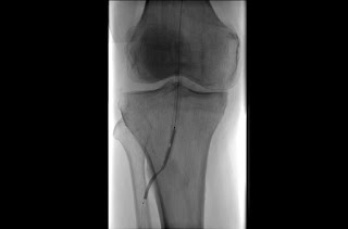
4. Angiography
This is a special type of X-ray technique for viewing blood
vessels and organs, especially the heart, by injecting a contrast agent into
the blood that enhances its visibility on the X-ray image.
figure 8: Angiography of a blood vessel in the region of the knee
5. Fluoroscopy
This is a type of medical imaging that produces a continuous
live X-ray image of the patient’s internal structure on a monitor.
Figure 9: The colon is clearly seen on the air-contrast barium enema
In security sector
X-rays are useful for checking
bags at airport check-ins. X-rays pass straight through soft materials such as
leather and plastic but are blocked by the metal in guns, knives and weapons.
Typically suitcases and bags travel up through large scanners on conveyor
belts, with X-ray images of their contents appearing instantly on computer
screen studied by security guards.
Figure 10: X-ray
use in the airport
In aeronautical engineering
X-ray help to track down things
like cracks and fatigue in metal components. Turbine blades in airplane jet
engines are tested in this way to make sure they are not harboring any problems
that would cause them to fail suddenly during flight.
Figure 11: turbine blade
In photography and art industry
Fine art black
and white floral photographic prints can be made from X-ray films of flowers.
Optimum technique for making the x-ray image would use unscreened fine-grain
industrial x-ray film covered by thin light-protective plastic. The film is exposed
at low kilo voltage for multiple seconds with floral material laying on the
light-protected film in specimen radiographic equipment. The film is hand
developed. Once the x-ray image is created, it can be used like any
photographic negative to make a print or digital image.
Figure 12: fine art black and white floral photographic print
And also X-ray imaging used in oil
paintings in order to prove the artist authenticity.
In scientific research area
X-ray is use in studying the inner
structure of materials. If fire beam of X-ray at a crystal, the atoms scatter
the beam in a very precise way, casting a kind of shadow of the crystal’s
interior pattern from which you can measure the distance between one atom and
its neighbors. This is called X-ray diffraction or X-ray crystallography.
Astronomy
X-ray used looking through
telescope to see light from distant objects even ones far out into space. Radio
telescopes, for example, are more like giant satellite dish antennas that
capture radio waves being given off from those distant sources. X-ray also
travel through space and we can study them in a similar way with telescope
tuned to recognize their particular frequency.
Contributed by
Felcons
W.M Madushan
A.M.N Chathurani
M.P.H Molagoda
Invaders
T.M.N.M Gunathilake
R.P.B.A Vitharana
S.A.S Lokuge
MSN
N.M.K Sarathchandra
R.M.P.N Rathnayake
S.M.S Siriwardena
Pixelz
S Jasintha
M.C.P Nifra
S Kishokanth
M.H.J.A Marrikkar
Scorpion
V.G.G.N Ariyarathna
N.P.J Withana
R.T.L Thennakoon


